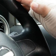
The forearm is made up of two bones in your lower arm, the radius and ulna. The forearm consists of two relatively parallel bones that connect two joints: elbow and wrist. A fracture in the forearm can occur near the wrist, in the middle of the forearm or near the elbow. The forearm motion allows us to rotate our palms up or down. A broken forearm can affect your ability to rotate your arm and even bend or straighten the wrist and elbow.
Radial Shaft Fractures
An isolated fracture of the radial shaft is an unusual injury. More commonly, fractures of the radial shaft are associated with injury to the ulna. When an isolated radial shaft fracture occurs, it commonly requires surgery unless the fracture is non-displaced.
Anatomy
The radius is the thicker and shorter of the two long bones in the forearm. It is located on the lateral side of the forearm parallel to the ulna (in anatomical position with arms hanging at the sides of the body, palms facing forward) between the thumb and the elbow.
Diagnosis
Diagnosis of a radial shaft fracture comes from a history, physical examination and lateral radiographs of the elbow, forearm and wrist. If there is the deformity of the radius, your physician will conduct immediate clinical diagnosis on the injury and will confirm the analysis with the result of the X-rays and will conduct a differential diagnosis that will include the scaphoid and the wrist because fractures of the radius are always accompanied by wrist fractures. If the fractures are not clearly seen on X-rays, delayed X-rays, MRI or CT scan will confirm the diagnosis further. Rehabilitation or rehab is necessary.
Treatment
Non-operative treatment of radial shaft fractures is rare and outcomes of nonsurgical treatment in the past have been poor. In the case of nondisplaced radial shaft fractures, initial immobilization in a splint will likely be the preferred treatment option. After swelling subsides, a long arm cast can be added.
Generally the standard of care for radial shaft fractures is open reduction and internal fixation. The surgical approach depends on the fracture location.
Complications
Early complications of radial head or neck fractures or dislocations include the following:
- Compartment syndrome
- Neurovascular injury
- Infection
Late complications include the following:
- Nonunion
- Hardware failure
- Malunion
- Infection
- Synostosis
- Persistent pain
Ulnar Shaft Fractures
An isolated fracture to the ulna often called a “nightstick” fracture, most often occurs during an altercation. Forearm fractures more commonly occur in men than women, owing to a higher incidence in men of participation in contact sports, altercations, falls from height, and motor vehicle collisions
Anatomy
The ulna is a long bone found in the forearm that stretches from the elbow to the smallest finger. When it is in the anatomical position, it is found on the medial side of the forearm. Ulna runs parallel to the radius and the other long bone in the forearm. The ulna is found to be slightly longer than the radius and thinner compared to the forearm and because the radius is thicker than the ulna, the radius is considered to be the larger one among the two.
The fracture of the ulna occurs as a result of fragility and normally from following a fall such as a fall on an outstretched hand. Special types of the broken ulna or the fractured ulna are:
Monteggia fracture-This occurs when there is a fracture of the proximal one-third of the ulnar shaft which is accompanied by the radial head.
Hume fracture occurs when there is a fracture of the olecranon accompanied by the anterior dislocation of the radial head.
Galeazzi’s fracture- At the distal radio-ulna junction, Galeazzi’s fracture occurs where the distal radius is fractured and the ulnar head dislocates.
Diagnosis
Standard AP, lateral, and oblique radiographs of the elbow and forearm should be obtained. It is important for the forearm views to also include the wrist. When assessing these views, normal radiographic lines should be evaluated to rule out injury.
Treatment
For nondisplaced or minimally displaced nightstick fractures, initial treatment consists of placement of a sugar-tong splint for 7-10 days. This can be followed by functional bracing for 8 weeks with active range of motion exercises for the elbow, wrist, and hand, or simple immobilization in a sling with a compression wrap, depending on the patient’s symptoms.
If the radial head cannot reduce despite ulna reduction and stabilization, the annular ligament, or rarely the radial nerve, may be interposed. Associated radial head fractures may require open reduction and fixation; and if so, the annular ligament should be repaired.
Complications
Although uncommon, persistent radial head instability can occur following anatomic reduction of the ulna.
Complications associated with Ulnar Shaft Fractures include:
- Damage to the nerves or blood vessels of the forearm
- Abnormal pressure build-up within the muscles of the arm/elbow that reduces blood flow, preventing oxygen and nourishment from reaching the nerve and muscle cells (termed as forearm compartment syndrome)
- The immobilization required to heal forearm fractures may occasionally result in painful shoulder inflammation, stiffness, and reduced range of motion (frozen shoulder)
- Malunion: A serious complication that might occur, if the ulnar bone in the forearm heals in an improper position
- Nonunion: Another serious complication, when the ulnar bone does not heal adequately, shifts to an improper position, receives an inefficient supply of blood flow, or becomes infected
- Osteoporosis: A disease that causes bones to weaken and become more brittle
- Infection of the bone (osteomyelitis)
- Reflex sympathetic dystrophy syndrome: A disorder associated with the sympathetic nervous system (a part of the autonomic nervous system) is a rare complication
Both Bones Forearm Fracture
Both bones fracture is an injury that almost always requires surgery in an adult patient. Without surgery, the forearm is generally unstable and there is no ability to cast this type of fracture. In younger children, nonsurgical treatment can be considered, but even in adolescents surgery may need to be performed. Both bones forearm fractures are most commonly treated by placing a metal plate and screws on both the radius and ulna bones.
Diagnosis
The forearm fractures are routinely diagnosed with the help of plain radiographs, including anteroposterior and lateral views of the forearm. A standard anteroposterior view of the forearm is taken with the elbow extended and the forearm in full supination.
Treatment
The ultimate goal of fracture management is to obtain acceptable reduction, stable fixation and bone healing, early return to activities of daily living, preservation of function and minimizing complications. Isolated ulna fractures have shown high success rates in non-operative management, including long-arm cast, functional brace, and close follow-up by serial x-rays.
The forearm fractures are usually managed by surgical methods, including closed or open reduction and internal fixation. Open reduction helps achieve anatomic reduction, stable fixation, and an early range of motion, thereby enabling patients to return to their pre-injured state as early as possible. Closed reduction and internal fixation can be performed using an intramedullary nail as a minimally invasive procedure which has a good favorable outcome in the pediatric population.
Complications of Forearm Fractures
The most common complications of these fractures include:
- Decreased Motion
Limited motion is common after the treatment of forearm fractures. Motion can be limited in the elbow and wrist joints but is most commonly noticed as a limitation of forearm rotation (i.e. opening a jar or turning a door handle).
- Non-Healing Fracture
The bones of the forearm can have inadequate healing leading to persistent pain. This is especially true with forearm fractures where bone is lost because of the type of fracture (i.e. many small pieces) or open fractures. Repeat surgery for bone grafting may be necessary in these cases.
- Infection
Infection can occur after any surgical procedure. When an infection occurs after fixation of a forearm fracture, the metal plate and screws may require removal in order to cure the infection.
- Painful Hardware
The metal implants used during surgery may be felt under the skin, and they may be painful. If they do cause discomfort they can be removed, usually at least a year after surgery.
Call Ventura Orthopedics Today!
Remember to check in with your body. If you are experiencing pain in your elbows and forearms or if you feel numbness or tingling in your hands and arms, it may be time to consult a medical professional.
The experienced and dedicated orthopedic surgeons at Ventura Orthopedics are here for you. If you are concerned about problems that you are experiencing with your elbows and forearms talk to the experts at Ventura Orthopedics today. Call us today at 800-698-1280 to schedule an appointment. With five convenient locations throughout Ventura County, California – in Ventura, Oxnard, Camarillo, Thousand Oaks, and Simi Valley, we are happy to help you get back in the game.


